Dr. Phil Zeltzman is a traveling, board-certified surgeon in Allentown, PA. His website is www.DrPhilZeltzman.com. He is the co-author of “Walk a Hound, Lose a Pound” (www.WalkaHound.com).
Kelly Serfas, a Certified Veterinary Technician in Bethlehem, PA, contributed to this article.
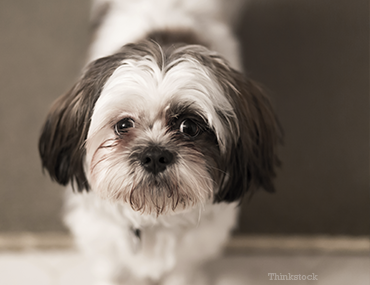
Moose, a 14 year old Shih Tzu, had been coughing more and more over the past few months. Not too concerned initially, his owners eventually took him to their family vet. Shockingly, chest X-rays showed a large mass in his left lung.
The vet referred Moose to our surgery service. After reviewing the X-rays and the blood work, I had a long heart-to-heart with Moose’s owners about the only definitive treatment: open chest surgery, which is considered a specialized surgery.
[Editor’s Note: Pet insurance is a great way to offset the cost of surgeries.]
Lung surgery
There are two ways to approach this type of surgery: either by cutting through the chest bone (a.k.a. sternum), or by cutting between two ribs, where only muscles are cut. Because Moose had a tumor in the left lung, the best approach was to go between two ribs on the left side. After surgery, Moose would need to be closely monitored at the hospital for a few days.
We discussed the possibility of taking a needle biopsy of the mass before surgery to learn more about the tumor, but agreed that it would not be very helpful.
Reasons included:
- The procedure would delay surgery by about a week, until the biopsy results were in.
- The surgery would be the same whether the mass was benign (abscess, pneumonia, benign tumor) or a malignant (a.k.a. cancerous) tumor.
- The mass was most likely cancerous.
- The client wanted the mass removed regardless of diagnosis. Moose’s guardians were determined to give him every chance.
Removing the mass from the lung
Each lung (left and right) is not a single sac, but made of several parts or lobes. After opening Moose's chest, we found an orange sized mass (in a 16 pound Shih Tzu!) in the left lower lung lobe. Removing it was not exactly easy, but it came out safely.
The next task was to place a device called a “chest tube,” which is a fairly large silicone tube, in the chest. It protrudes from the body to allow the nurses to remove air or fluid which may build up after this type of surgery.
The chest was closed up, and Moose was recovered in ICU. There, he received pain medications and antibiotics, as well as plenty of TLC.
Recovering from lung surgery
Moose recovered smoothly. The next morning, he was surprisingly bright and alert. He even jumped into my arms when I went to check on him! We aspirated the chest tube with a syringe every few hours. The amount of fluid and air decreased every time we did this. When the amount became small enough, we removed the chest tube.
Moose was kept under close observation for another 12 hours, to make sure no problems occurred after removal of the chest tube. He was then sent home.
I went over the discharge instructions with his guardians; they basically entailed pain medications and antibiotics for 1 week, a lovely plastic cone around his neck for 2 weeks, and strict confinement for a month.
Confirmation of lung cancer
About a week later our suspicion was confirmed: the tumor was an adenocarcinoma, which is a type of lung cancer. The bad news would have been neither chemotherapy nor radiation therapy would have helped much. The good news was we had removed the mass entirely.
I had another discussion with Moose’s guardian, and we agreed that we should track his progress closely by scheduling follow up exams every two months. Little Moose has been doing great ever since the surgery.
If you have any questions or concerns, you should always visit or call your veterinarian -- they are your best resource to ensure the health and well-being of your pets.
